Why Choose Us?
-

Quick turn around
We get reports to you in a timely manner. Our goal is a turnaround time of one to five hours
-

Expert Reports
Well qualified and experienced team providing you with highly detailed and professional interpretation
-

Affordable pricing
Get your dental radiology reports at an affordable fees
-

Consultation
Looking for a second opinion or just a consultation? You’re invited to send radiographs our way for consultation at any time
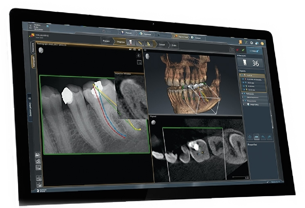
Cone Beam Computed Tomography
Cone beam computed tomography (CBCT) is a radiographic imaging method that allows accurate, three-dimensional (3D) imaging of hard tissue structures. CBCT is the most significant among the medical diagnostic imaging modalities that have emerged recently.
This imaging modality is capable of providing sub-millimeter resolution (2 line pair/mm) images of higher diagnostic quality, with shorter scanning times (~60 s). Radiation exposure dose from CBCT is 10 times less than from conventional CT scans during maxillofacial exposure (68 µSv compared with 600 µSv of conventional CT) and also it has got great dimensional accuracy (only about 2% magnification).
Increasing availability of this technology is now providing the dental clinician an imaging modality, which is capable of providing a 3D representation of the maxillofacial structures with minimal distortion and reduced radiation hazards.
At Dental Scans and Reports we provide full radiological reporting of your scans and also virtual face to face discussion of reports.
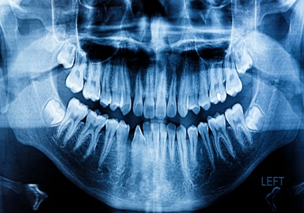
2D Dental Panoramic Tomography
The Dental Panoramic Tomogram (OPG) is a must have for every dental patient because it provides an overview of the entire jaw and all the teeth and surrounding structures on one film.
What Is a Panoramic X-Ray?
A panoramic x-ray is a two-dimensional x-ray that captures a picture of a patient’s entire mouth in just one image. The x-ray includes a view of all of the teeth, multiple bones of the head and neck, and other critical anatomical structures.
Unlike bitewing dental x-rays which show a close up of view of your teeth, this x-ray gives a global view of a patient’s head and neck. This view allows for diagnosing more than just regular dental concerns, like cavities or gum disease. Rather, this view enables the radiologist to see other important issues that may be occurring in the surrounding tissue and jaw bones like oral cancer or other abnormalities, things that would not be visible in an ordinary dental radiograph.
Intraoral Views
Bitewing X-rays show details of the upper and lower teeth in one area of the mouth. Each bitewing shows a tooth from its crown (the exposed surface) to the level of the supporting bone. Bitewing X-rays detect decay between teeth and changes in the thickness of bone caused by gum disease.
The Bitewing examination is the most widely taken radiological exam globally. It is the most critical means of detecting even the smallest caries in between the teeth.
For every Dental Practitioner, it provides a means of recording and documenting the restorative treatment being undertaken as well as those which will be needed in the future. medical insurance companies rely on submission of the Bitewing to confirm approve restorative treatment or to confirm past treatment claims.
Sialogram
A sialogram is an x-ray test using contrast (x-ray dye) to look in detail at the larger salivary glands (the parotid or submandibular). These glands help to keep your mouth moist by draining saliva into your mouth through small tubes called ducts.
This examination (Sialography) is done by introducing a very thin tube into the opening of the duct and injecting a small amount of contrast (Dye). This probe procedure is done without any anesthesia since it is not painful.
A sialogram is performed for patients suspected of having a blockage of the salivary flow due to stones or strictures.
The signs and symptoms of salivary gland blockage include, swelling of one or more glands; pain and or swelling of a gland before meals among others.